Inside Rodin’s Hands
April 17, 2014
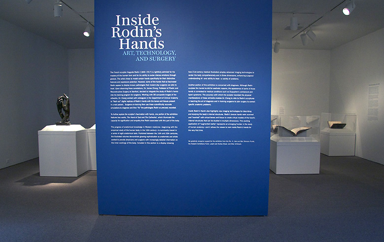
Over the past year I’ve been helping the Cantor Arts Center organize an exhibition titled Inside Rodin’s Hands: Art, Technology, and Surgery which opened last week on the Stanford campus. The show features the French sculptor Auguste Rodin’s sculpted hands that display or resemble known pathologies that modern-day surgeons are able to treat. The exhibit also traces the history of anatomical knowledge and visualization from the 16th century to the present day. I contributed by writing gallery text about the historical anatomical books on display and designing the show’s graphics.
The exhibit was organized by Dr. James Chang, Professor of Plastic and Reconstructive Surgery at Stanford, who integrates the study of Rodin’s hands into his training program for surgeons. In his undergraduate seminar, “Surgical Anatomy of the Hand: From Rodin to Reconstruction,” students study the hands that Rodin created and work with 3D digital simulations of those artworks to diagnose and “fix” the pathologies that Rodin recorded. In pairing art and science, Dr. Chang aims to make anatomy lessons uniquely engaging while connecting humanities students with medicine and acquainting premeds with the arts.
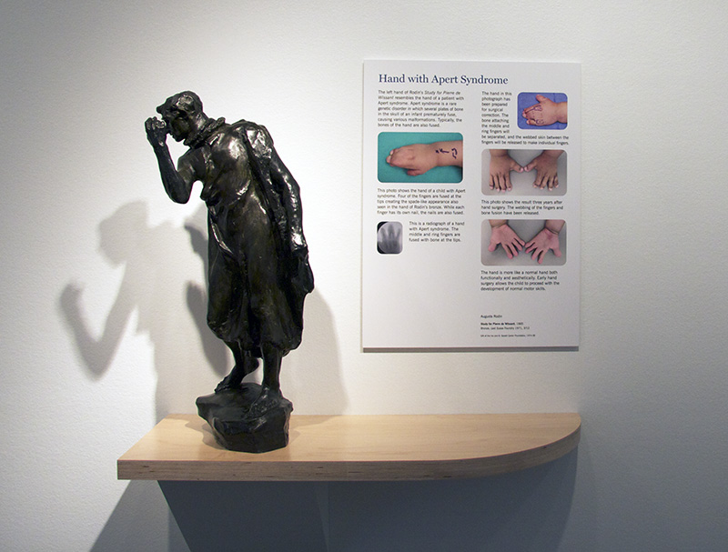
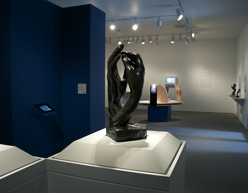
There are three sections in the exhibition. The first is concerned with diagnosis and contains Rodin sculptures accompanied by photos of real patients’ hands with the same condition. The text and pictures illustrate contemporary surgical procedures such as realigning fingers using plates and screws, removing a ganglion cyst with exacting surgery, reattaching a severed thumb, and even substituting a big toe for a lost thumb.
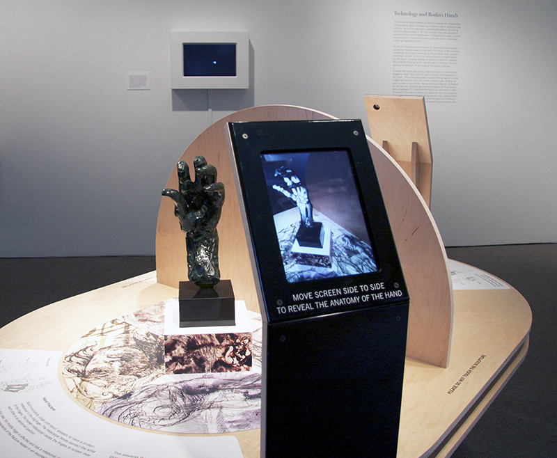
The next portion of the exhibition is interactive; visitors move an iPad in an arc around Rodin’s bronze hands and see computer-generated graphics of the bones, nerves, and muscles that might be seen within actual hands. This display describes new imaging technologies that provide three-dimensional views and a sense of depth into internal structures — an application of “augmented reality” representing an emerging frontier in the study of human anatomy.
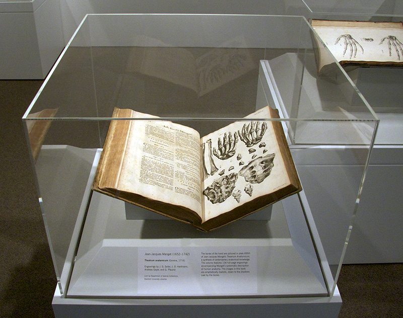
The final section consists of a series of eight anatomical texts chronicling the progress of Western medicine beginning with the empirical study of the human body in the 16th century. Ranging in publication from 1585 to 1834, the illustrated volumes demonstrate growing attention by anatomists and artists to providing physicians and surgeons with increasingly accurate information on the inner workings of the body.
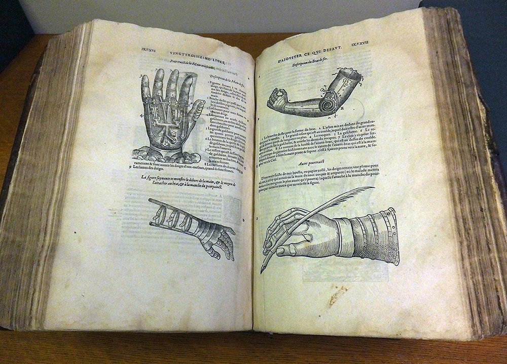
The books also reveal actual and proposed medical practices of the day. One of my favorites on display is Les Oeuvres d’Abroise Paré, pictured above. Published in Paris in 1585, it compiles material from Ambroise Paré’s 26 books, ranging from stories of sea monsters to drawings of artificial limbs. Paré contributed greatly to both the practice of surgical amputation and the invention of scientific instruments. He introduced the implantation of teeth, limb prostheses, and artificial eyes made of gold and silver. In this spread Paré addresses the subject of the hand with inventively depicted mechanical appendages, human muscles manipulated with pulleys, and related surgical instruments.
Inside Rodin’s Hands is on view through August 3, 2014. The Cantor Arts Center is open Wednesday through Sunday, 11am – 5pm, Thursdays until 8pm; admission is free.